MRI Safety Information for Advanced Bionics Cochlear Implants
For Italy
Testing has demonstrated that the Advanced Bionics HiRes implant family is MRI Conditional. Conditions vary by geography. A patient with the implant may be safely scanned with MRI only under very specific conditions. Scanning under different conditions may result in severe patient injury.
MRI Safety Information by Type of Implant
| Type of Implant | MRI Field Strength (T) |
|---|---|
| C1.0* | MRI is contraindicated |
| C1.2* | MRI is contraindicated |
| CII* | MRI is contraindicated |
| HiRes 90K* | 1.5T |
| HiRes 90K Advantage | 1.5T |
| HiRes Ultra w/magnet | 1.5T |
| HiRes Ultra magnet removed | 3.0T** |
*These devices are no longer sold in the EU
**For MRI the magnet has to be removed surgically

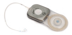
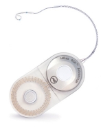
HiRes™ Ultra Cochlear Implant
MRI information
| Device | HiRes Ultra |
|---|---|
| Instructions For Use | HiResolution Bionic Ear System: HiRes Ultra |
| 1.5T magnet removed | Scanning ok under certain conditions |
| 1.5T magnet in place with MRI Antenna Coil Cover and bandaging protocol CI-7521 | Scanning ok under certain conditions |
| 3.0T magnet removed | Scanning ok under certain conditions |
1.5T with the magnet in place
MRI Warning
Do not allow patients with a HiRes Ultra cochlear implant to be in the area of an MRI scanner unless the following conditions have been met:
The bandaging protocol [Refer to the MRI Antenna Coil Cover (CI-7521) IFU for details] recommended by Advanced Bionics is followed when the patient undergoes an MRI procedure with the magnet left in place, or
For cases that would clinically benefit from reduced device artifact (for example some head or neck scans), the internal magnet is surgically removed and possibly replaced with the temporary nonmagnetic plug before the patient undergoes an MRI procedure.
The external sound processor and headpiece are MR Unsafe and must be removed before entering a room containing an MR scanner.
The recommended minimum duration of time post implant surgery prior to undergoing an MRI scan is 2 to 4 weeks in order to allow any inflammation to subside.
An MRI scan is not recommended if the patient has a fever.
For additional information regarding the use of an MRI scanner with a HiRes Ultra device, please contact Advanced Bionics Technical Support.
For 1.5T MRI scans with the magnet in place
- Horizontal closed bore scanners with a static magnetic field of 1.5T (magnet in place).
- Maximum MR system reported, whole body averaged specific absorption rate (SAR) of ≤ 2 W/kg at 1.5T for quadrature transmit RF body coils.
- Maximum MR system reported, head averaged SAR of ≤ 3.2 W/kg at 1.5T for quadrature transmit RF head coils.
- RMS gradient field of 30 T/s and peak gradient field of 150 T/s.
- Maximum spatial field gradient of 3.47T/m at 1.5T (magnet in place)
When tested under the scan conditions defined above the HiRes Ultra implant produced a maximum temperature of < 3° after 15 minutes of continuous 1.5T Scanning.
In MRI testing of unilateral recipient conditions, the image artifact caused by the device extends from the HiRes Ultra implant approximately 7.9 cm in a 1.5T MRI with the magnet in place.
In MRI testing of bilateral recipient conditions >9.5 cm in a 1.5T MRI with the magnet in place using a spin echo or gradient echo pulse sequence.
These artifacts may result in a loss of diagnostic information in the implant vicinity.
Please note that the MRI Antenna Coil Cover CI-7521 and bandaging supplies must be on hand at the time of the MRI procedure.
1.5T with the magnet removed
MRI Warning
Do not allow patients with a HiRes Ultra cochlear implant to be in the area of an MRI scanner unless the following conditions have been met:
The bandaging protocol [Refer to the MRI Antenna Coil Cover (CI-7521) IFU for details] recommended by Advanced Bionics is followed when the patient undergoes an MRI procedure with the magnet left in place, or
For cases that would clinically benefit from reduced device artifact (for example some head or neck scans), the internal magnet is surgically removed and possibly replaced with the temporary nonmagnetic plug before the patient undergoes an MRI procedure.
The external sound processor and headpiece are MR Unsafe and must be removed before entering a room containing an MR scanner.
The recommended minimum duration of time post implant surgery prior to undergoing an MRI scan is 2 to 4 weeks in order to allow any inflammation to subside.
An MRI scan is not recommended if the patient has a fever.
For additional information regarding the use of an MRI scanner with a HiRes Ultra device, please contact Advanced Bionics Technical Support.
For 1.5T MRI scans with the magnet removed
- Horizontal closed bore scanners with a static magnetic field of 1.5T (magnet removed).
- Maximum MR system reported, whole body averaged specific absorption rate (SAR) of ≤ 2 W/kg at 1.5T for quadrature transmit RF body coils.
- Maximum MR system reported, head averaged SAR of ≤ 3.2 W/kg at 1.5T for quadrature transmit RF head coils.
- RMS gradient field of 30 T/s and peak gradient field of 150 T/s.
- Maximum spatial field gradient of 13.90T/m at 1.5T with magnet removed
When tested under the scan conditions defined above the HiRes Ultra implant produced a maximum temperature of < 3° after 15 minutes of continuous 1.5T Scanning.
In MRI testing of unilateral recipient conditions, the image artifact caused by the device extends from the HiRes Ultra implant approximately 3.1 cm in a 1.5T MRI with the temporary non-magnetic plug and In MRI testing of bilateral recipient conditions 4.2 cm in a 1.5T MRI with the temporary non-magnetic plug using a spin echo or gradient echo pulse sequence.
These artifacts may result in a loss of diagnostic information in the implant vicinity.
Please note that the MRI Antenna Coil Cover CI-7521 and bandaging supplies must be on hand at the time of the MRI procedure.
Magnet Removal/Replacement Using the HiRes Ultra Magnet Tool Kit, CI-1418
These tools must be sterilized prior to use. See the ”Guide for Reprocessing HiRes Ultra Reusable Tools” provided in the kit.
3.0T with the magnet removed
MRI Warning
The internal magnet is surgically removed and possibly replaced with the temporary non-magnetic plug before the patient undergoes an MRI procedure.
Do not allow patients with a HiRes Ultra cochlear implant to be in the area of an MRI scanner unless the following conditions have been met:
The bandaging protocol [Refer to the MRI Antenna Coil Cover (CI-7521) IFU for details] recommended by Advanced Bionics is followed when the patient undergoes an MRI procedure with the magnet left in place, or
For cases that would clinically benefit from reduced device artifact (for example some head or neck scans), the internal magnet is surgically removed and possibly replaced with the temporary nonmagnetic plug before the patient undergoes an MRI procedure.
The external sound processor and headpiece are MR Unsafe and must be removed before entering a room containing an MR scanner.
The recommended minimum duration of time post implant surgery prior to undergoing an MRI scan is 2 to 4 weeks in order to allow any inflammation to subside.
An MRI scan is not recommended if the patient has a fever.
For additional information regarding the use of an MRI scanner with a HiRes Ultra device, please contact Advanced Bionics Technical Support.
3.0T MRI scans with the magnet removed
- Horizontal closed bore scanners with a static magnetic field of 3.0T (magnet removed)
- Maximum MR system reported, whole body averaged specific absorption rate (SAR) of ≤ 2 W/kg at 3.0T for quadrature transmit RF body coils.
- Maximum MR system reported, head averaged SAR of ≤ 2.6 W/kg at 3.0T for quadrature transmit RF head coils.
- RMS gradient field of 30 T/s and peak gradient field of 150 T/s.
- Maximum spatial field gradient of 6.90T/m at 3.0T with magnet removed
When tested under the scan conditions defined above the HiRes Ultra implant produced a maximum temperature of < 3° after 15 minutes of continuous 1.5T Scanning.
In MRI testing of unilateral recipient conditions, the image artifact caused by the device extends from the HiRes Ultra implant approximately 4.7 cm in a 3.0T MRI with the temporary non-magnetic plug.
In MRI testing of bilateral recipient conditions 4.7 cm in a 3.0T MRI with the temporary non-magnetic plug using a spin echo or gradient echo pulse sequence.
These artifacts may result in a loss of diagnostic information in the implant vicinity.
Please note that the MRI Antenna Coil Cover CI-7521 and bandaging supplies must be on hand at the time of the MRI procedure.
Magnet Removal/Replacement Using the HiRes Ultra Magnet Tool Kit, CI-1418
These tools must be sterilized prior to use. See the ”Guide for Reprocessing HiRes Ultra Reusable Tools” provided in the kit.
HiRes™ 90k Cochlear Implant Family
MRI Information
| Device | HiRes 90k and HiRes 90K Advantage |
|---|---|
| Instructions For Use | HiResolution Bionic Ear System: HiRes 90K and 90K Advantage |
| 1.5T magnet removed | Scanning ok under certain conditions |
| 1.5T magnet in place with MRI Antenna Coil Cover and bandaging protocol CI-7521 | Scanning ok under certain conditions |
MRI Warning
The HiRes 90K implant family, with either the internal magnet removed or left in place, has been tested with 1.5 Tesla/64 MHz and 0.3 Tesla/12 MHz MRI systems.
MRI is contraindicated except under the circumstances described below.
Do not allow patients with an implant from the HiRes 90K family to be in the area of an MRI system unless:
- The bandaging protocol recommended by Advanced Bionics (029-M486) is followed when the patient undergoes an MRI procedure with the magnet left in place or,
- The internal magnet is surgically removed and possibly replaced with the Magnet Insert Dummy before the patient undergoes an MRI procedure.
- The external sound processor and headpiece are removed before entering a room where an MRI scanner is located.
MRI parameters should be selected to ensure a Specific Absorption Rate (SAR) of less than 1.0 W/kg in the head region.
Patient should be monitored verbally and visually throughout the MRI procedure.
Image shadowing may extend as far as 70 cm2 (without magnet) and 210 cm2 (with magnet) around the implant, resulting in loss of diagnostic information in the implant vicinity. The extent of the shadowing may be minimized by adjusting the signal parameters.
For additional information regarding the use of MRI, please read the Magnetic Resonance Imaging (MRI) for the HiRes 90K and HiRes 90K Advantage CI (9050050269) or contact Advanced Bionics Technical Support.
Note: In the EU, MRI Examination Request Form (029-M569) must be provided to Advanced Bionics for review and approval prior to performing the MRI.